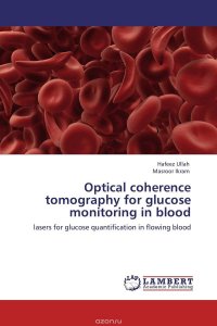
Optical coherence tomography (OCT) is an emerging biomedical imaging technique with very high spatial resolution. It relies on the coherence properties of lasers, to generate micron-scale cross sectional subsurface images of tissue. Clinically, several uses of this imaging method appear attractive; for example, We have implemented OCT in glucose quantification of blood in vitro and in vivo in research lab. OCT’s ability to quantify blood flow dynamics in-vivo also opens up several exciting possibilities to study diseases with significant involvement of microcirculation; for example, we have quantified the measured changes in blood flow (in vitro) during and following higher concentrations and in vivo scenario by envisioning the blood microvasculature with the help of speckle variance optical coherence tomography. Brownian motion of the scatterers is affected due to presence of glucose as measured by OCT. The temporal analysis of Brownian motion statistics yields the translational diffusion coefficient and viscosity of non-flowing and flowing fluids. The increases in glucose concentrations deform red blood cells resulting in rouleaux formations;thus, affecting the OCT signal. Это и многое другое вы найдете в книге Optical coherence tomography for glucose monitoring in blood
Optical coherence tomography for glucose monitoring in blood
Подробная информация о книге «Optical coherence tomography for glucose monitoring in blood ». Сайт не предоставляет возможности читать онлайн или скачать бесплатно книгу «Optical coherence tomography for glucose monitoring in blood »
